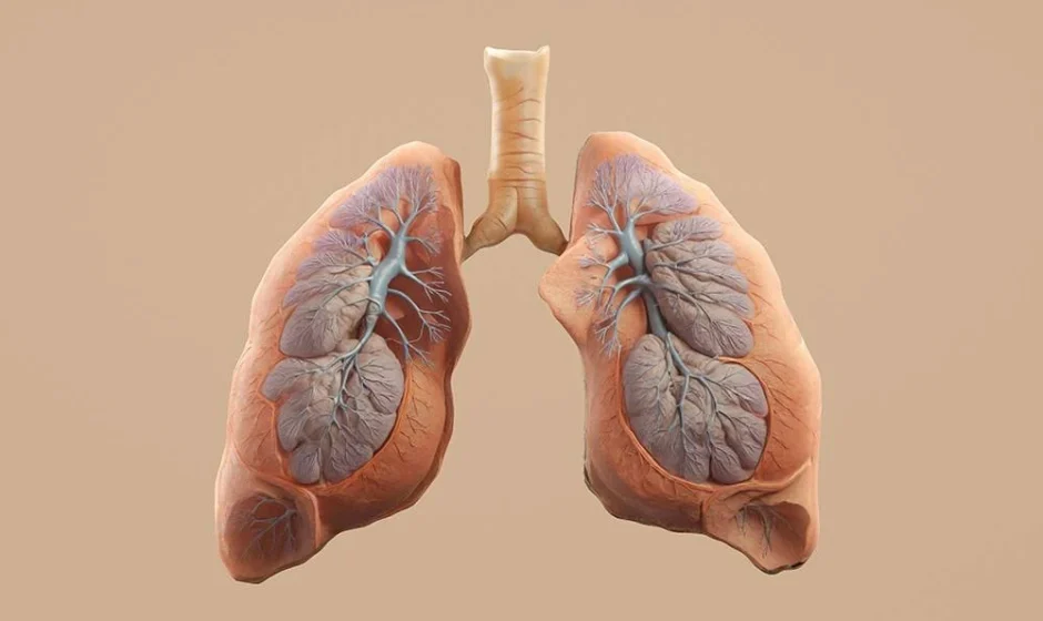

A collapsed lung sounds alarming, and in many cases, it absolutely is. Medically known as pneumothorax, this condition occurs when air leaks into the space between the lung and the chest wall, causing part or all of the lung to collapse. While some cases resolve with minimal intervention, others can become life-threatening within minutes. Understanding the different types of pneumothorax, what causes them, and how they present clinically is essential for recognizing the condition early and responding appropriately.
What Happens During a Pneumothorax
The lungs are surrounded by a thin double-layered membrane called the pleura, with a small amount of fluid between the layers that allows smooth movement during breathing. When air enters this pleural space, normal negative pressure is disrupted, and the lung can no longer expand properly. The result is reduced oxygen exchange, shortness of breath, and in severe cases, cardiovascular compromise. The severity of symptoms often depends on how much air has accumulated and how quickly the condition develops.
Spontaneous Pneumothorax
Spontaneous pneumothorax occurs without any obvious external trauma and is divided into two subtypes. Primary spontaneous pneumothorax happens in individuals without known underlying lung disease, often tall, thin young adults, and is typically linked to small air-filled blisters called blebs that rupture on the lung surface. Secondary spontaneous pneumothorax occurs in people who already have lung disease, such as chronic obstructive pulmonary disease, cystic fibrosis, or tuberculosis, where weakened lung tissue is more prone to rupture. Secondary cases tend to be more dangerous because the affected individual often has less pulmonary reserve to begin with.
Traumatic Pneumothorax
Traumatic pneumothorax results from injury to the chest, such as a stabbing, gunshot wound, rib fracture, or blunt force trauma from a motor vehicle accident. The injury creates an opening that allows air to enter the pleural space, either from outside the body or from a punctured lung. Because trauma patients frequently have multiple injuries occurring simultaneously, traumatic pneumothorax can be easy to miss without a thorough chest assessment and imaging.
Iatrogenic Pneumothorax
Iatrogenic pneumothorax is caused by a medical procedure rather than an external injury. Common culprits include central line placement, thoracentesis, lung biopsy, and positive pressure mechanical ventilation. Because so many hospitalized patients undergo these procedures, iatrogenic pneumothorax is more common than many people realize, making post-procedure monitoring an important part of patient safety protocols.
Tension Pneumothorax
Tension pneumothorax is the most dangerous form and represents a true medical emergency. It occurs when air continues to enter the pleural space with each breath but cannot escape, creating a one-way valve effect. As pressure builds, it not only collapses the affected lung but also pushes the mediastinum, including the heart and great vessels, toward the opposite side of the chest. This shift can compress the unaffected lung and impede venous return to the heart, leading to severe hypotension, distended neck veins, tracheal deviation, and rapid cardiovascular collapse if not treated immediately.
Recognizing the Symptoms
While symptoms vary by type and severity, common signs of pneumothorax include sudden, sharp chest pain that often worsens with breathing, shortness of breath, rapid breathing, rapid heart rate, and decreased or absent breath sounds on the affected side. In tension pneumothorax specifically, patients may show signs of shock, including pale or bluish skin, severe anxiety or confusion, and a rapidly dropping blood pressure. Because these symptoms can escalate quickly, prompt recognition is critical to preventing further deterioration.
Diagnosis and Treatment Approaches
Diagnosis typically involves a physical examination combined with imaging, most commonly a chest X-ray or bedside ultrasound, though tension pneumothorax is often a clinical diagnosis made before imaging can be obtained due to the urgency of the situation. Treatment depends on severity. Small, stable pneumothoraces may be managed with observation and supplemental oxygen, allowing the air to reabsorb naturally. Larger or symptomatic cases often require needle decompression or placement of a chest tube to remove trapped air and allow the lung to re-expand. Tension pneumothorax requires immediate needle decompression, typically followed by chest tube placement, as delaying treatment even briefly can be fatal.
Why Clinical Knowledge of Pneumothorax Matters
Because pneumothorax can present in such varied ways, from a vague ache in a young athlete to sudden cardiovascular collapse in a trauma patient, clinicians benefit from a strong working knowledge of how each type develops and progresses. Recognizing subtle early signs, understanding which patients are at higher risk, and knowing when a situation has escalated to an emergency are skills built through both experience and structured education. Professionals looking to reinforce this knowledge often turn to courses that provide a focused clinical review of pneumothorax types and management, reinforcing the assessment skills needed to catch deterioration early.
Final Thoughts
Pneumothorax is a condition that spans the spectrum from minor and self-limiting to immediately life-threatening, and the difference often comes down to type, underlying health status, and how quickly it is recognized. Staying current on these distinctions is not just academic; it is a direct contributor to faster recognition and better patient outcomes. Nurses and respiratory therapists seeking to strengthen their clinical assessment skills can explore pneumothorax-focused nursing CEUs as a way to earn continuing education units while building the knowledge needed to catch deterioration early. Earning CEUs in critical care topics like tension pneumothorax ensures that time spent on professional development translates directly into sharper bedside judgment.